
FIG. 3.
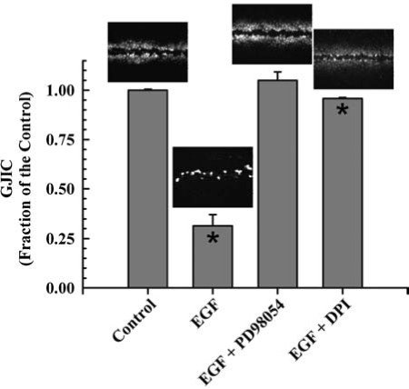
Involvement of Mek and NADPH oxidase in EGF-induced inhibition of GJIC. The figure was graphed with data obtained from ref. 101 with permission of Acta Medica Nagasakiensia. Averaged results of the scrape load-dye transfer data with a representative image of the cells at a magnification of 200X is placed above each bar. The concentrations of epidermal growth factor (EGF), diphenyleneiodonium (DPI), and PD98059 were 10 μg/l, 5 μM, and 20 μM respectively. Cells were incubated for 15 min with EGF and preincubated with PD98059 and DPI for 15 and 20 min, respectively. Quantitative results were from triplicate sets of data of SL/DT images presented. Only the EGF and EGF + DPI treated cells were significant (*) from the control (ANOVA, p < 0.001, F = 304; Tukey post-hoc p < 0.05). The cells used for these experiments is the established epithelial cell line F344 WB isolated from the livers of adult male Fischer 344 rats (100) generously donated from Drs. J. W. Grisham and M. S. Tsao, University of North Carolina (Chapel Hill, NC).