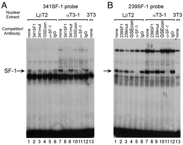
Fig. 5. SF-1 Interacts with the FSHβ Promoter at Two Sites.

EMSA was performed using a probe spanning either the −341 SF-1 element (A) or the −239 SF-1 element (B) along with nuclear extracts from LβT2 (lanes 1–6), αT3-1 (lanes 7–12), or NIH3T3 (lane 13) cells. Competitions were performed using 100-fold excess unlabeled oligonucleotide as indicated above each lane: None (lanes 1, 7, and 13; no competitor), 341SF1 (A, lanes 2 and 8; wild-type 341SF-1 element; 5′-TTGGTTTACCTTCGCAATGGAG-3′), 341mut (A, lanes 3 and 9; mutated 341SF-1 element; 5′-TTGGTTTAAATTCGCAATGGAG -3′), 239SF1 (B, lanes 2 and 8; wild-type 239SF-1 element; 5′-TTTAATTTACAAGGTGAGGGAG-3′), 239mut (B, lanes 3 and 9; mutated 239SF-1 element; 5′-TTTAATTTAGAATTTGAGGGAG-3′), GSEcon (lanes 4 and 10, consensus GSE from the α-GSU promoter; 5′-GCTGCTGACCTTGTCACTAGCT-3′). The GSE is underlined and the mutated sequences are shown in bold. Antibodies directed against SF-1 (α-SF-1) or normal rabbit IgG (IgG) were included in the reactions as indicated. Arrow indicates the SF-1 containing complex.
