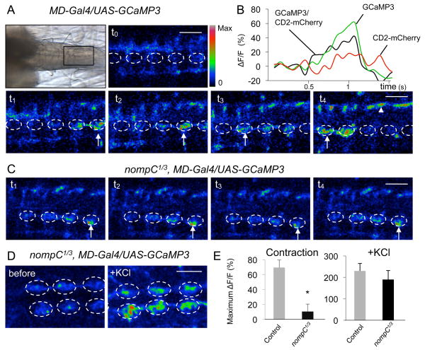
Figure 2. NompC is required for the activation of MD neurons during peristaltic muscle contractions.
(A) Ca2+ response of MD neurons to peristaltic muscle contractions in control larvae. Top left panel, a transmitted light image showing the ventral nerve cord in a larval preparation. Anterior is to the left and posterior to the right. (t0 to t4) Time-lapse images of Ca2+ signals (arrows) generated by spontaneous muscle contractions. The dorsalmedial neuropil is outlined with a dashed circle. The nerve bundles are marked with arrowheads. Color scale indicates activation level (red is the highest). Scale bar, 30 μm.
(B) Representative fluorescence change (ΔF/F) of GCaMP3 and CD2-mCherry (internal reference control) in control larvae.
(C) Ca2+ response to muscle contractions in nompC1/3 mutant larvae. Scale bar, 30 μm.
(D) Representative images before and after adding KCl to nompC1/3 mutant larvae. Scale bar, 50 μm.
(E) Quantification of GCaMP responses. Left panel, responses to muscle contractions for control (n=8) and nompC1/3 larvae (n=10). Right panel, responses to KCl for control (n=3) and nompC1/3 larvae (n=3). The GCaMP signal is normalized to CD2-mCherry reference signal. * p<0.01. See also Movie S2.

