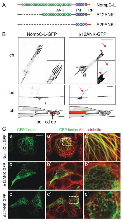
Figure 5. Ankyrin repeats of NompC are required for ciliary localization and microtubule association.
(A) A schematic of full-length and truncated NompC-L protein structures. ANK, ankyrin domain; TM, transmembrane domain; TRP, TRP box domain.
(B) Localization of NompC-L-GFP and Δ12ANK-GFP in chordotonal organs (upper panels) and bd neurons (middle panels). The lch5 sensory neurons are indicated by *, the base of the cilium by an arrowhead and the ciliary tip by an arrow. The bd axon is indicated by an arrow. A schematic interpretation of the localization of NompC-L-GFP and Δ12ANK-GFP in chordotonal organs are shown in the bottom panels. pc, proximal cilium; cd, ciliary dilation; dc, distal ciliary tip. Scale bars, 20 μm.
(C) Localization of NompC-L-GFP, Δ12ANK-GFP and Δ29ANK-GFP in HEK 293 cells. Full-length and truncated NompC-L protein was visualized by the GFP fluorescence (green). The cells were co-stained with anti-α-tubulin (red). Boxed area in a′, b′, c′ is enlarged and shown in a″, b″, c″ respectively. Scale bar, 10 μm. See also Figure S4.

