
Figure 2.
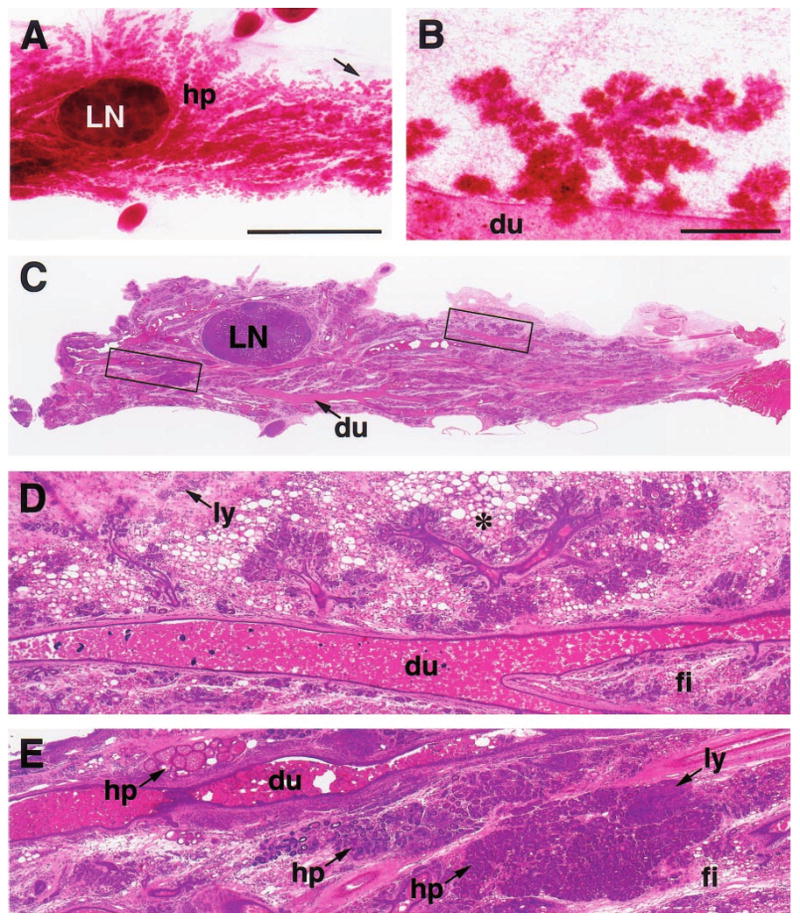
Carmine-stained wholemount (a,b) and H&E-stained paraffin section (c – e) of an abdominal (#4) mammary gland with diffuse hyperplasia (hp), fibrosis (fi) and lymphocytic infiltration (ly) from a 15-month-old parous WAP-Str1 mouse sacrificed 4 months after its pups were removed. The hyperplastic branches indicated by the arrow in (a) are outlined in (c) and are shown at higher magnification in (b and d). These sparse and disproportionately short secondary branches terminate in relatively well-developed lobuloalveolar structures and are surrounded by multilocular adipocytes (asterisk). The boxed area to the left of the central lymph node (LN) in c is enlarged in e and shows three hyperplastic areas, each with a distinct histologic appearance. Dilated (ectatic) primary ducts (du) containing considerable amounts of residual secretory material are also evident throughout the gland. Scale bars, 5 mm (a,c), 500 μm (b,d,e)