
Figure 5.
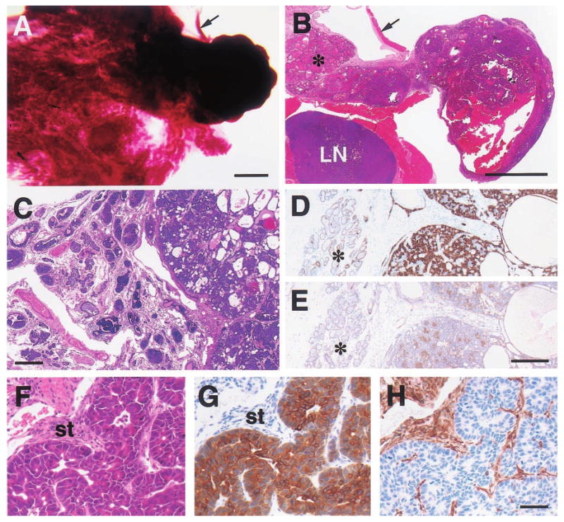
Histologic appearance of moderately well-differentiated mammary adenocarcinomas from 15- (a,b), 19- (c – e) and 23-month-old (f – h) WAP-Str1 transgenic mice as seen after wholemount (a), H&E (b,c,f), anti-cytokeratin-8 (d,g), anti-smooth muscle actin (e) and anti-vimentin (h) staining. (a,b) The tumor at right contains cystic spaces and necrotic debris, and sits adjacent to a diffuse lactational-like hyperplasia (asterisk) and a lymph node (LN) that is surrounded by muscle. The strip of connective tissue and skeletal muscle (arrow) is indicated for orientation. (c – e) This complex tumor (at right in each panel) contains numerous cystic spaces and tumor cell nests composed of mixed small and large cell populations. The small cells are smooth muscle actin-positive (e) and have small nuclei with a dense chromatin structure. The larger cells are cytokeratin-8 positive (d), have larger nuclei with a more open chromatin structure, and exhibit distinct intercellular bridges. The tumor is surrounded by a fibrotic stroma, atypical papillary lesions (c) and areas of secretory hyperplasia (asterisk). (f – h) These serial sections show a papillary adenocarcinoma with large cytokeratin-positive tumor cells, numerous mitotic figures, and an abundant vimentin-positive stroma (st). Scale bars, 2 mm (a,b), 300 μm (c), 200 μm (d,e), 50 μm (f – h)