
Fig. 1.
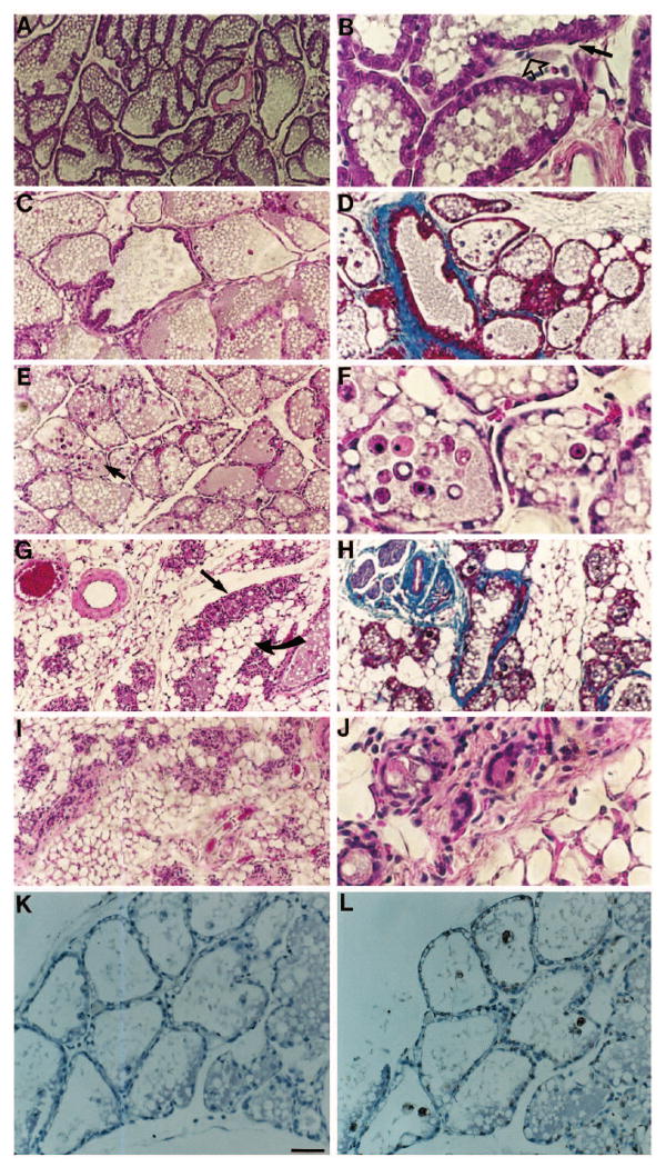
Hematoxylin-eosin and trichrome staining of lactating and involuting mouse mammary gland. (A,B) Mammary gland after 7 days of lactation. Open arrow indicates fibroblasts; solid arrow indicates myoepithelial cells. (C,D) Mammary gland after 2 days of involution. (E) Mammary gland after 3 days of involution. Arrow indicates apoptotic cells in the lumen of an alveolus shown in higher magnification in F. (G,H) Mammary gland after 4 days of involution. Straight arrow indicates a group of collapsed alveoli; curved arrow indicates adipocytes. (I,J) Mammary gland after 8 days of involution. (K,L) Staining for apoptotic cells after 3 days of involution. (K) control without and (L) with terminal deoxynucleotidyl transferase added. A,C,D,E,G,H,I, bar, 100 μm; B,F,J,K,L, bar, 25 μm. A,B,C,E,G,I,J were stained with hematoxylin and eosin; K,L were counterstained with hematoxylin; D,H were stained with trichrome.