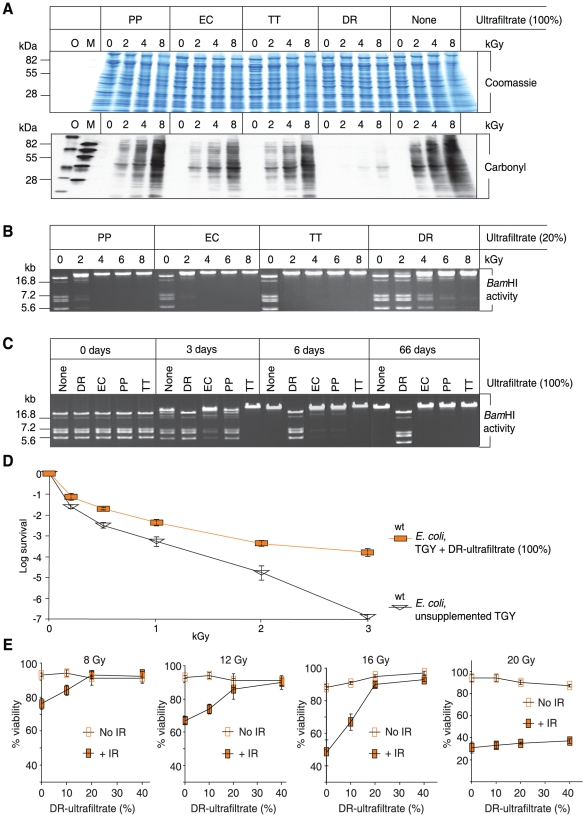
Figure 1. In vitro and ex vivo protection by DR-ultrafiltrate.
(A) DR-ultrafiltrate prevents protein oxidation. The indicated ultrafiltrates were mixed with purified E. coli proteins and irradiated to the indicated doses of μ-radiation (kGy). Proteins were then separated by polyacrylamide gel electrophoresis and visualized by Coomassie staining. Duplicate gels were subjected to Western blot carbonyl analysis, which reveals the presence (black) or absence (no signal) of protein oxidation. PP, P. putida; EC, E. coli; TT, T. thermophilus; and DR, D. radiodurans. O and M, size-standards. (B) DR-ultrafiltrate preserves the activity of an irradiated enzyme. BamHI was irradiated in the indicated ultrafiltrates, then incubated with μ-DNA and subjected to agarose gel electrophoresis. (C) DR-ultrafiltrate preserves the activity of a desiccated enzyme. BamHI was desiccated from the indicated ultrafiltrates and stored in a desiccator for the indicated times, and then assayed for residual activity as in panel B. (D) DR-ultrafiltrate protects E. coli. Wild-type E. coli (MM1925) cells were grown in TGY medium supplemented with DR-ultrafiltrate and irradiated without change of broth to the indicated doses, then recovered on TGY medium. Colony forming unit (CFU) survival assays were in triplicate for each dose, with standard deviations shown. (E) DR-ultrafiltrate protects human Jurkat T cells. DR-ultrafiltrate was added to the growth medium 1 day before irradiation. The viability of irradiated cells was determined by trypan blue staining 2 days after irradiation. Viability assays were in triplicate, with standard deviations shown.

