Figure 3.

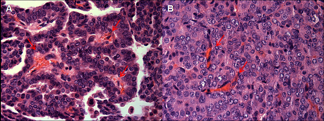
A. Early mouse lung adenoma with papillary structures showing prominent central vascular core, designated by arrows. B. Advanced mouse lung adenoma with solid tumor growth pattern and disorganized vascular network, designated by arrows.

A. Early mouse lung adenoma with papillary structures showing prominent central vascular core, designated by arrows. B. Advanced mouse lung adenoma with solid tumor growth pattern and disorganized vascular network, designated by arrows.