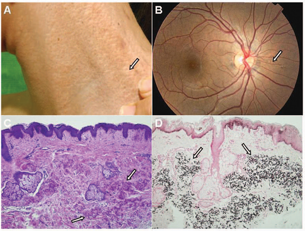
Figure 1.
Diagnosis of PXE in the proband of the family. Note the characteristic cutaneous lesions on the side of the neck (A) and angioid streaks in the eye (B) (arrow). Histopathology reveals accumulation of mineralized elastotic material in mid-dermis when examined by Hematoxylin and Eosin (C) or von Kossa (D) stains (arrows).

