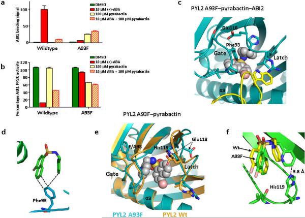
Figure 5.
A93F converts PYL2 into a pyrabactin-activated receptor. (a+b) PYL1 A93F is activated by pyrabactin to bind and inhibit ABI1 as determined by AlphaScreen assay (a) and by phosphatase assay (b); (n=3, error bars=s.d.). (c) Close view of pyrabactin in the PYL2 A93F–pyrabactin–ABI2 trimeric complex. ABI2 with Trp290 is shown in yellow. (d) Phe93 interacts with both ring systems of pyrabactin in the PYL2 A93F–pyrabactin–ABI2 structure. (e) Structure of pyrabactin in the PYL2 A93F ligand binding pocket (cyan) overlaid with the PYL2 wildtype structure (brown). (f) His119 is flipped into the ligand binding pocket to allow direct interaction with the pyridine ring of pyrabactin in the PYL2 A93F–pyrabactin structure.

