
Figure 3.
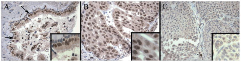
Tissue micro-arrays (TMAs) of nuclear staining for BRMS1 in normal bronchial epithelium and human lung cancer specimens. (A) TMA of human normal bronchial epithelium (arrows) stained with BRMS1 antibody, as described in Materials and methods, demonstrates strong and diffuse nuclear immunoreactivity. (B) TMA of human lung adenocarcinoma tissue stained with BRMS1 antibody, as described in Materials and methods, demonstrates moderate, diffuse nuclear immunoreactivity. (C) TMA of human lung squamous cell carcinoma tissue stained with BRMS1 antibody, as described in Materials and methods, demonstrates an absence of staining.