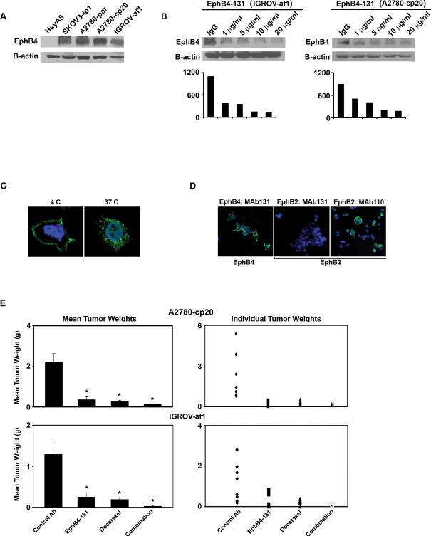
Figure 1. Targeting EphB4 with EphB4-131 monoclonal antibody in ovarian cancer cell lines.
A) Western blot analysis of EphB4 expression in ovarian cancer cell lines. B) A2780-cp20 and IGROV-af1 cells were treated by indicated amount of EphB4-131 antibody for 48 hours. Cells were then lysed and subjected to Western blot analysis to assess EphB4 expression. Corresponding densitometry graphs are included. C) Biotinylated EphB4-131 was localized in A2780-cp20 cells using streptavidin-FITC. Confocal images were taken at 100x. EphB4 bound MAb131 (green) was internalized at 37 °C only. D) EphB4-131 specific staining was seen in cells expressing EphB4 (left panel) but not in cells expressing EphB2 only (middle and right panel). EphB2 specific antibody MAb 110 was used as a positive control. Nuclei were counterstained with DAPI (blue). E) EphB4-131 alone and in combination with docetaxel significantly decreased tumor growth in A2780-cp20 and IGROV-af1 tumor models (*p<0.05 compared to control antibody group). An unrelated control antibody was used as negative control.

