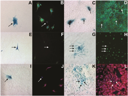
Figure 1.
Bright field (A, C, E, G, I, K) and fluorescent microscopy (B, D, F, H, J, L) images demonstrating MDSCs differentiation into hepatocyte-like cells when co-cultured with the AML12 hepatocyte cell line after 1 day (A, B, E, F, I, J) and 7 days (C, D, G, H, K, L). In the bright field images, MDSCs are denoted by β-galactosidase expression (blue) and by black arrows whereas in fluorescent images, they are denoted by white arrows at the same location. In the fluorescent images, the co-cultured cells are stained for albumin (green: B, D), HNF4α (green: F, H) and αFP (red: J, L). The cell nuclei were stained with DAPI (blue).

