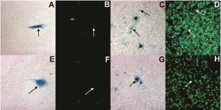
Figure 2.
Bright field (A, C, E, G) and fluorescent microscopy (B, D, F, H) images demonstrating MDSCs differentiation into hepatocyte-like cells when co-cultured with the Hepa1-6 hepatocyte cell line after 1 day (A, B, E, F) and 7 days (C, D, G, H). MDSCs are denoted by β-galactosidase expression (blue) and black arrows in bright field microscopy images. Fluorescent images, were taken at the exact location, where MDSCs are denoted by white arrows and stained for albumin (green: B, D) and HNF4α (red: F, H). The cell nuclei were stained with DAPI (blue).

