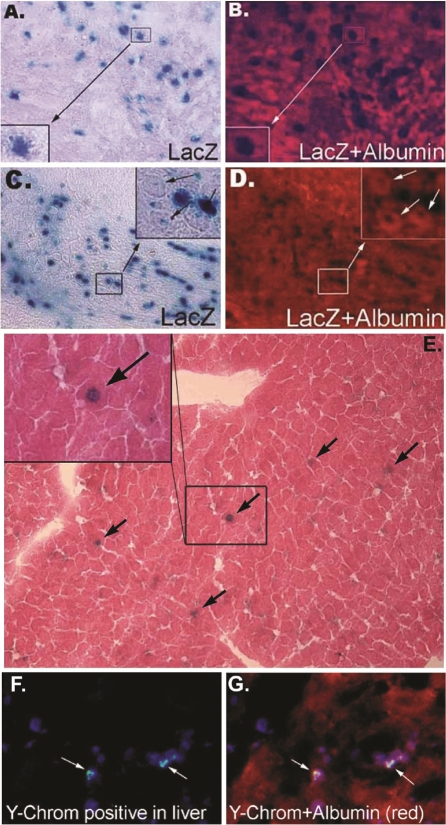
Figure 4.
MDSCs differentiate into liver-like cells in vivo. Images of liver tissue harvested 3 days after MDSCs transplantation and stained for β-galactosidase (A, blue) or albumin (B, red). Similarly, liver tissue harvested 7 days after MDSC transplantation was also stained for β-galactosidase (C, blue) or albumin (D, red). Long term MDSCs engraftment into hepatectomized livers was observed after 3 months (E). As indicated by the black arrows, β-galactosidase positive (blue) MDSCs are found in the liver tissue subjected to hemotoxylin and eosin staining. A magnified image is shown in the inset of this figure. FISH analysis revealing the donor signal (Ychromosome) within injured liver tissue of female recipients, 7 days after male MDSCs were transplanted (F, G). MDSCs were stained for cell nuclei, blue; Y-chromosome, green; and albumin, red.

