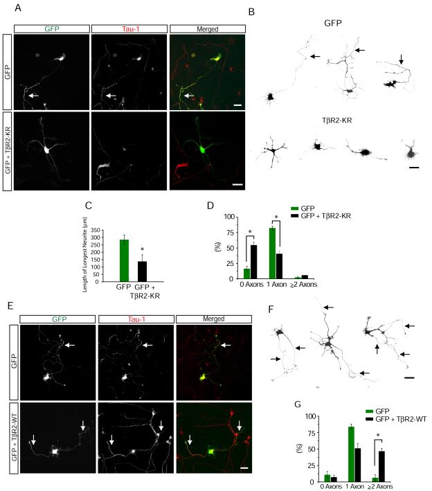
Figure 4.
Cell Autonomous TGF-β Signaling Mediates Axon Formation
(A) Dissociated hippocampal neurons from E18 rat embryos expressing GFP or GFP + TβR2-KR were fixed and stained for the axonal marker tau-1. Cells were transfected 4-6 hours after plating and fixed 65-72 hours later. Arrow indicates tau-1 positive axons, which are absent from cells expressing TβR2-KR. Scale bar, 20 μm.
(B) Camera lucida traces of neurons expressing GFP (top) or GFP + TβR2-KR (bottom). Arrows indicate axons. Scale bar, 20 μm.
(C) Data represent means ± SEM of the longest neurite in control cells and cells expressing TβR2-KR. n = 12, *p<0.05, Student’s t-test.
(D) Quantification of axon number in cells expressing GFP alone or GFP + TβR2-KR. Data pooled from at least 3 independent experiments. GFP, n = 42; TβR2-KR, n = 53; *p<0.05, Student’s t-test.
(E) Neurons expressing GFP or GFP + TβR2-WT showing multiple tau-1 positive axons emerging from cells expressing TβR2-WT (arrows). Scale bar, 20 μm.
(F) Camera lucida traces of neurons expressing TβR2-WT. Arrows indicate axons. Scale bar, 20 μm.
(G) Quantification of axon number in cells expressing GFP alone or GFP plus TβR2-WT. GFP, n = 41; TβR2-WT, n = 44; *p<0.05, Student’s t-test.

