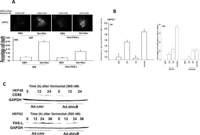
Figure 6. Vorinostat promotes sorafenib toxicity by increasing expression of FAS-L.
Panel A. Upper IHC: HEPG2 cells plated in 8 well chamber slides were pre-treated with control IgG or an IgG to neutralize FAS-L (1 μg/ml) and 30 min later treated with vehicle (DMSO) or sorafenib (3 μM), and vorinostat (500 nM) in combination. Cells were fixed 6h after exposure and surface levels of CD95 determined by IHC. The density of CD95 staining was determined in 40 cells (n = 2, +/- SEM). Lower Graph: HEPG2 cells were pre-treated with control IgG or an IgG to neutralize FAS-L (1 μg/ml) and 30 min later treated with vehicle (DMSO) or sorafenib (3 μM), and vorinostat (500 nM) in combination. Forty eight h later cells were isolated and viability determined by trypan blue exclusion (n = 3, +/- SEM). Panel B. Left Graph: HEPG2 cells 24h after plating in 96 well plates were transfected with NFκB-luciferase and β-galactosidase constitutive reporter constructs. Thirty six hours after transfection cells were treated with vehicle (DMSO), sorafenib (3.0 μM), vorinostat (500 nM) or both sorafenib and vorinostat. Cells were assayed for NFκB-luciferase and β-galactosidase activity 24h after treatment (± SEM, a representative from 2 separate studies). Control studies demonstrated that over-expression of dominant negative IκB blocked vorinostat-induced activation of NFκB-luciferase activity (not shown). Right Graph: HEPG2 cells 24h after plating were infected with control empty vector virus (CMV) or a recombinant virus to express dominant negative IκB S32A S36A (dn IκB). Twenty four hours after infection, cells were treated with vehicle (DMSO), sorafenib (3.0 μM), vorinostat (500 nM) or both sorafenib and vorinostat. Forty eight hours after drug exposure, cells were isolated, spun onto glass slides and stained using established methods for double stranded DNA breaks indicative of apoptosis (TUNEL) as described in the Methods (n = 2, +/- SEM). Panel C. HEPG2 and HEP3B cells 24h after plating were infected with control empty vector virus (CMV) or a recombinant virus to express dominant negative IκB S32A S36A (dn IκB). Twenty four hours after infection, cells were treated with vehicle (DMSO) or vorinostat (500 nM). Cells were isolated 12h and 24h after treatment. SDS PAGE and immunoblotting were performed to determine changes in the expression of CD95 and FAS-L, as indicated. A representative is shown of 3 separate studies.

