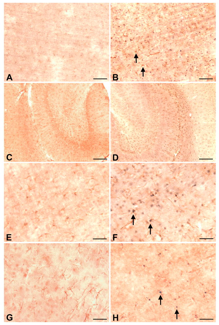
Figure 4. MCAO induces reactive gliosis and nuclear translocation of STAT3 in astrocytes.
At 24 h following MCAO in young rats, enhanced immuonstaining of GFAP-positive cells (astrocytes) was observed in ipsilateral cortex (B) compared to the GFAP immunostaining observed in contralateral cortex (A). Basal GFAP immunostaining was observed in contralateral hippocampus (C) that was enhanced in iplateral hippocampus (D). MCAO also resulted in the appearance of STAT3 immunoreactivity in astrocytic nuclei (B, D, F). High magnification microscopy reveals the lack of STAT3 immunoreactivity in astrocytes of the contralateral cortex (E), while the nuclei of astrocytes in ipsilateral cortex appear blue-black from double-immunocytochemical labeling (F). At 24 h following MCAO in aged rats, enhanced GFAP and STAT3 immunostaining also was observed in ipsilateral cortex (H) compared to GFAP and STAT3 immunostaining observed in contralateral cortex. In general GFAP and STAT3 immunostaining in ipsilateral cortex of aged rats (H) was less than that observed in young rats (F). Arrows denote STAT3 immunostaining in GFAP-positive astrocytes. Bar A - B = 100 μ, C - D = 200 μm, E - H = 50 μ.

