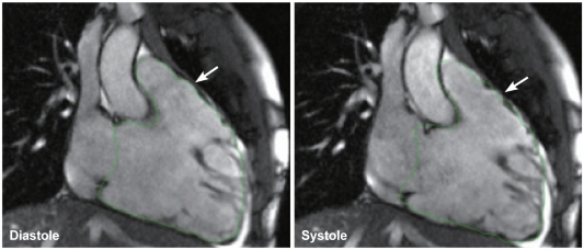
Fig. 8.
Cine images in 2-chamber view of the right ventricle show right ventricular dilatation, poor systolic function, and dyskinesia of the anterior wall of the right ventricular outflow tract (arrow). The right ventricular contour was drawn on the diastolic image as shown in the left panel. The contour was copied and overlain on the systolic image as shown in the right panel. Note the minimal downward excursion of the base of the right ventricle in systole. The anterior wall of the right ventricular outflow tract shows protrusion in systole beyond the limit of right ventricular contour drawn on the diastolic image.

