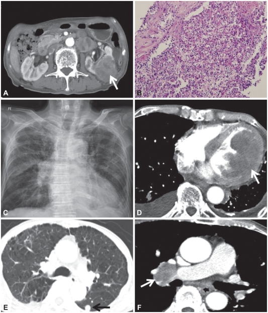
Fig. 1.
Renal cell carcinoma (RCC) with left ventricular and pulmonary metastases, but without right ventricular metastasis. A: abdominal computed tomography (CT) shows a very large RCC in the left kidney (white arrow) with thickening of the renal fascia. B: pathology obtained from the renal biopsy shows a typical clear cell renal cell carcinoma (H&E stain, ×200). C: plain chest radiograph shows cardiomegaly, right hilar enlargement, and reticular opacities in both lungs. D: chest CT shows a heterogenous mass lesion (white arrow) involving the entire left ventricle without right ventricular metastasis. E: multiple pulmonary nodules consistent with pulmonary metastases. Note underlying lung emphysema. F: a large metastatic lymph node in the right hilar area (white arrow).

