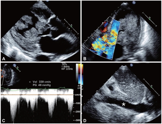
Fig. 2.
Two-dimensional (2D) echocardiography of metastasis to the left ventricle (LV) from renal cell carcinoma, with no metastasis in the inferior vena cava (IVC). (A) 2D echocardiogram shows a very large, lobulated, oscillating mass attached to the lateral wall of the LV and occupying the cavity of the LV (B), and (C) color flow Doppler showing turbulent jet and continuous-wave Doppler velocity through the left ventricular outflow tract (LVOT). (D) No evidence of mass in IVC (*).

