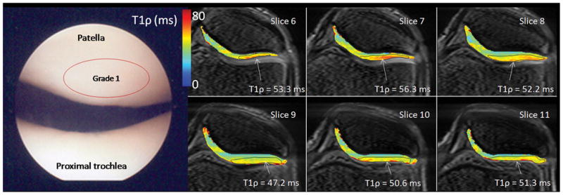
Figure 3.

Arthroscopic photographs and T1ρ relaxation maps from a 40 year old male (patient 1). The patient was observed at arthroscopy to have diffuse grade 1 chondromalacia throughout the entire knee joint. No focal defects or thinning was observed in either patellar or femorotibial compartments on MRI, however, a heterogeneous T1ρ distribution was observed, as well as local elevated T1ρ and cartilage thinning was observed in the lateral patellar superficial compartment and elevated T1ρ = 47.2–56.3 across six slices. Diffuse, low femoral condyle compartment T1ρ was observed (T1ρ = 38.2 ms).
