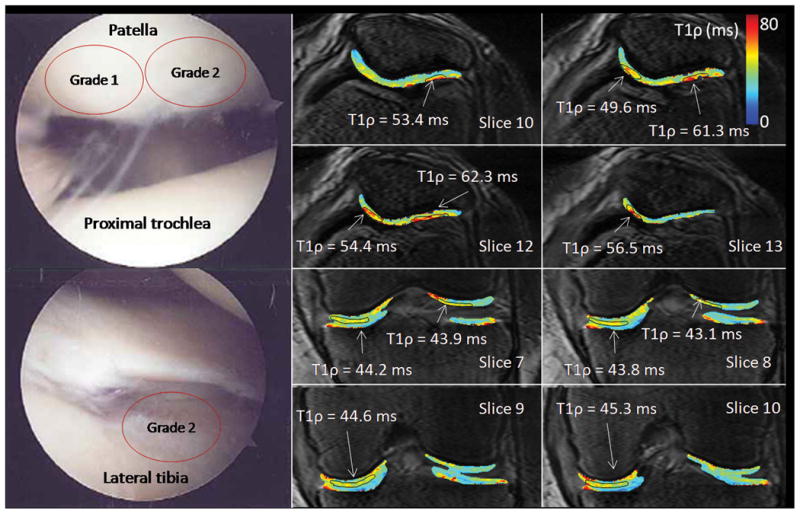
Figure 5.

Arthroscopic photographs and T1ρ relaxation maps from a 76 year old female (patient 6). This patient was observed at arthroscopy to have grade 1 chondromalacia of the lateral patellar facet and grade 2 chondromalacia of the medial patellar facet. At MRI, both the medial and lateral patellar facets had focal T1ρ lesions (T1ρ = 49.6–56.5 and 53.4–62.3 ms, respectively). Grade 2 chondromalacia was observed in the medial femorotibial compartment at arthroscopy and elevated T1ρ = 43.8–45.3 ms was observed laterally.
