
FIG. 4.
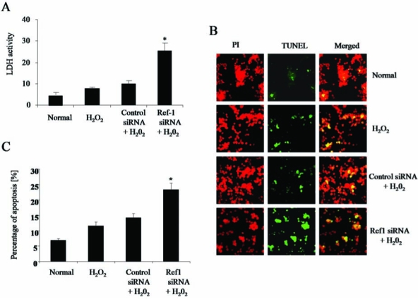
Ref-1 inhibition induces cell death. Adult cardiac stem cells were treated with either control or Ref-1 siRNA followed by H2O2 (10 μM), as mentioned in Methods. (A) The release of lactate dehydrogenase (LDH) enzyme from the cells was measured by using the spent culture medium obtained at the end of experimental period. (B) Fluorescent microscopic images showing the TUNEL staining of apoptotic cells (green channel) and nucleus (red channel, propidium iodide). (C) Quantification of apoptotic cells stained with TUNEL. The results are expressed in percentages. PI, propidium iodide. (For interpretation of the references to color in this figure legend, the reader is referred to the web version of this article at www.liebertonline.com/ars).