Figure 3.
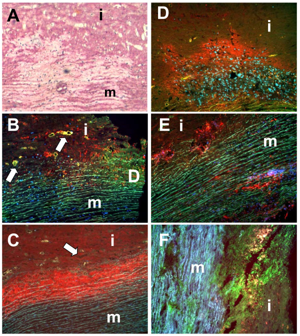
Correlation between inflammatory infiltration and collagen degradation. Picro Siriusred staining (panel A: collagen deposition = red) showing areas with massive collagen degradation. Immunohistochemistry for CD68 (panel B) and 27E10 (panel C) demonstrated accumulation of infiltrating cells particularly at the intima-media border (arrows indicate areas with profound vascularization). MMP1 (panel D) and MMP9 (panel E) were strongly expressed at the intima-media border and in deeper areas of the media. Interestingly, MMP2 expression was only very low (not shown). In situ zymography (panel F: collagenolysis) showed massive collagenolytic activity at the intima-media border and in deeper areas of the media. (m: media; i: intima; original magnification: ×100)

