FIG. 4.
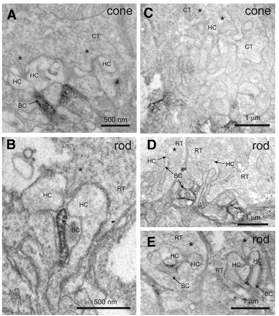
EYFP-nyctalopin is localized to the postsynaptic membrane of bipolar dendrites. Electron micrographs show staining for mGluR6 (A and B) and EYFP-nyctalopin (C–E) in nob rescue mice. Antibodies to mGluR6 stain invaginating processes of cone (A) and rod (B) photoreceptors. Antibodies to GFP stain EYFP-nyctalopin in nob rescue mice in or near cone (C) and rod (D and E) terminals. The staining of EYFP-nyctalopin is closely associated with cell membranes. Staining near cone terminals does not appear to involve the invaginating tip of the bipolar cells, rather it is slightly distal, at the base of the invagination. RT, rod terminal; CT, cone terminal, HC, horizontal cell dendrite; BC, bipolar cell dendrite; *, synaptic ribbon in rod and cone terminals.

