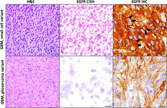
Figure 1.
Small cell GBMs are highly cellular tumors composed of monomorphic cells with ovoid nuclei. Most small cell GBMs harbor EGFR gene amplification, shown here by chromogenic in situ hybridization as innumerable red dots corresponding to EGFR gene copies in the tumor cell nuclei. EGFR gene amplification is typically associated with strong cell membrane staining (arrows) for the EGFR protein by IHC, although diffuse cytoplasmic staining is also seen. Gliosarcomas, which are characterized by a collagen-rich stroma, usually do not exhibit EGFR gene amplification. Moderate cytoplasmic EGFR immunopositivity may be seen even in tumors without EGFR gene amplification, but distinct cell membrane staining is rarely, if ever, present.

