Figure 1.
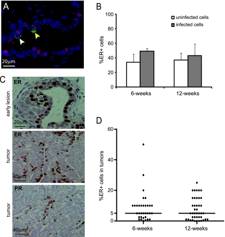
RCAS-mediated gene expression is detected in both ER+ and ER- mammary epithelial cells in MMTV-tva mice; RCAS-PyMT leads to ER+ mammary tumors in most infected MMTV-tva mice. (A and B) Pubertal (n = 4; 6 weeks) and adult (n = 9; 12 weeks) MMTV-tva mice were infected with RCAS-β-actin (HA-tagged) and killed 4 days later. Coimmunofluorescent staining for HA (green) and ER (red) was performed on the infected glands. A merged image that includes the DAPI nuclear counterstain is shown for an infected mammary gland from a 6-week-old mouse (A). A costained cell is indicated by a yellow arrow, and a non-costained cell is indicated by a white arrow. Bar graph (B) shows the percentage of ER+ cells in the general epithelium or among the infected cells (identified by staining for HA). (C) Six-week-old MMTV-tva mice (n = 5) were infected with RCAS-PyMT and killed 4 days later. Immunohistochemical staining for the indicated proteins was performed on the resulting early lesions as well as 33 tumors induced by infecting 6-week-oldmice with RCAS-PyMT (107 IU). (D) Dot plot showing the percentage of ER+ cells in these tumors as well as in tumors (n = 41) generated by intraductal injection of RCAS-PyMT (107 IU) into 12-week-old MMTV-tva mice. Horizontal bar indicates the median value.

