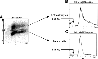
Figure 1.
Flow cytometry protocol to analyze the tumor cell death. The FITC-propidium iodide protocol was used to analyze the cell cycle of tumor cells (nonlabeled) and astrocytes (GFP-labeled) separately. The sub-G0 and -G1 regions were defined as the percentage of cell death. (A) The GFP gate is able to distinguish tumor cells (bottom) from astrocytes (top). (B) The cell cycle of astrocytes (FITC-positive because of GFP labeling). (C) The cell cycle of tumor cells (FITC-negative).

