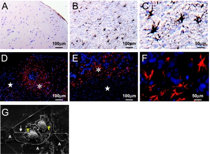
Figure 3.
Melanoma-astrocyte interaction. (A–C) A human specimen of melanoma brain metastasis in a tumor free zone (A) shows only sporadic expression of GFAP (brown); in the tumor zone (B), many reactive astrocytes surround and infiltrate tumor lesions; (C) higher magnification of reactive astrocytes in the tumor zone. (D–F) Mouse model of brain metastasis produced by human metastatic melanoma cell lines A375P (D) and TXM13 (E, F). Reactive astrocytes are stained in red, and nuclei are stained in blue. Asterisk (*) indicates tumor zone; star (⋆), tumor-free zone. (F) Higher magnification of reactive astrocytes from (E). (G) Scanning electron microscopic image of astrocytes (A) and A375P melanoma cells (T) shows direct physical contact between the astrocytes (extending pods-feet) and tumor cells. Note the seamless structure between the astrocytes and tumor cells indicated by the arrowhead.

