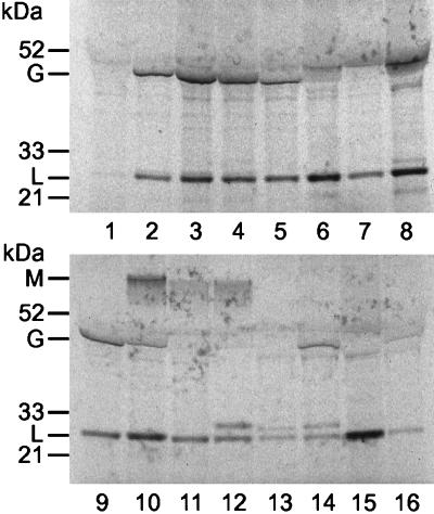
Figure 1.
Analysis of Ig secreted by the Karpas 707H cell line and hybridomas. The cells were grown at a concentration of 2 × 10⁶ cells per milliliter in l-methionine, l-cysteine-deficient medium that was supplemented with 10% dialyzed FBS and with [35S]methionine and [35S]cysteine at 250 μCi/ml (Amersham Pharmacia). The cells were incubated for 8 h at 37°C in a CO2 incubator. After incubation, the cell suspensions were centrifuged at 1,000 × g for 5 min. The supernatant was analyzed by SDS-PAGE after total reduction with 10% Bis-Tris Gel with Mops running buffer (Invitrogen NuPage Electrophoresis system). Tracks: 1, small quantity of light chain produced by the Karpas 707H cells; 2, IgG that reacts with the gp41 HIV-1 produced by the hybrid with the EBV-infected 164 cells; 3 and 4, IgG-producing hybridoma formed with fresh WBC; 5–9 and 13–16, hybridoma formed with tonsil cells. Track 10 may contain both IgG and IgM, probably because of a mixture of two hybridomas that were formed in the same well. Tracks 11 and 12, IgM-secreting hybridomas; tracks 12–14, hybridomas that secrete two distinct light chains. This figure illustrates that in such hybridomas the secretion of the myeloma λ light chain is greatly amplified compared with the nonfused myeloma (track 1). What appears to be a single line of light chains in most hybridomas is probably due to myeloma and donor chains banding in the same position.

