
Figure 3.
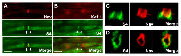
Localization of S4 in nodal regions of the PNS. Sections through rat sciatic nerve at p30 were labeled with antibodies to S4 (A-D, green) and Na+ (Nav, A, C-D, red) or K+ (Kv1.1, B, red) channels. Superposition of the labels is shown (merge). The paranodal regions are not labeled with antibodies against S4 (A, B, arrowheads). On sections perpendicular to nerve axis (D), S4 immunoreactivity is peripheral to Nav labeling. C, D: confocal stacks. Scale bars: 10 μm (A, B), 1 μm (C, D).