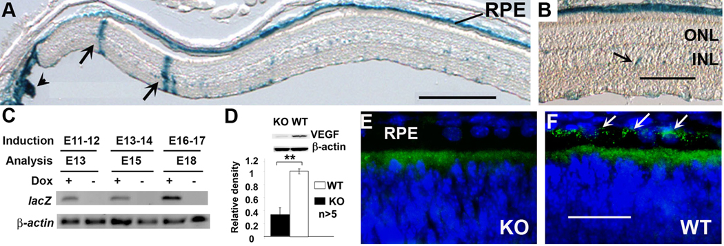
Figure 1.
Generation of conditional VEGF KO mice. A–B: localization of Cre expression in retinal sections of VRC2/R26R mice. Arrows in A: expression in Müller cells. Arrow head: expression outside the retina. Arrows in B: expression in unidentified cells in inner nuclear layer (INL). ONL: outer nuclear layer. Scale bars in A and B equal to 250 µm and 100 µm, respectively. C: RT-PCR analysis of Cre activated lacZ expression (30 cycles) in the retina of VRC2/R26R mice one day after dox induction at E11-12, E13-14, or E16-17. β-Actin (20 cycles) served as an internal control. D–F: Analysis of VEGF expression in P5 conditional VEGF knockout mice with immunoblotting and immunohistochemistry after dox induction at E16-18. Scale bar in F equals to 40 µm. **: p<0.001 vs WT in t-test. RPE-derived VEGF was expressed near the basolateral side of the RPE (towards choroid, arrows in F) in WT mice and was disrupted in the conditional VEGF KO mice.

