Figure 2.
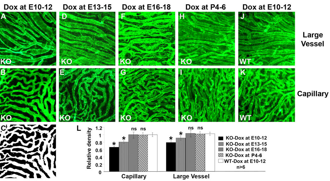
Temporal requirement of the RPE-derived VEGF in choroidal vascular development. A–K: representative images of anti-CD31 antibody-stained large choroidal vessels and capillaries from P21 conditional VEGF KO mice and WT mice after dox induction at E10-12, E13-15, E16-18, or P4–P6. C: black-white image converted from B, which was used to calculate the area of vessels. M: quantification of choroidal vascular density. *: p<0.05 vs WT in t-test. ns: not significant. Inducible disruption of the RPE-derived VEGF at various times resulted in regulatable changes in choroidal vascular density and the RPE-derived VEGF was required for the development of choroidal vasculature before embryonic day 15.

