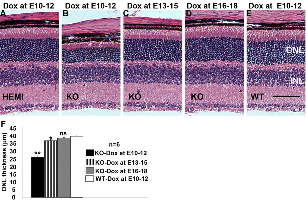
Figure 4.
Retinal morphology in P21 conditional VEGF KO mice after dox induction at various time for two days. A–E: H & E stained P21 retinal sections from homozygous and hemizygous (HEMI) conditional VEGF KO mice and WT controls with dox induction at E10-12, E13-15, or E16-18. ONL: outer nuclear layer. INL: inner nuclear layer. Scale bar: 40 µm. F: Average ONL thickness 0.5 mm from optic nerve. * and **: p<0.05 and p<0.001, respectively. ns: not significant. Inducible disruption of the RPE-derived VEGF at various times resulted in regulatable loss in rod photoreceptor function and photoreceptor ONL thickness in P21 conditional VEGF KO mice with dox induction before E15.

