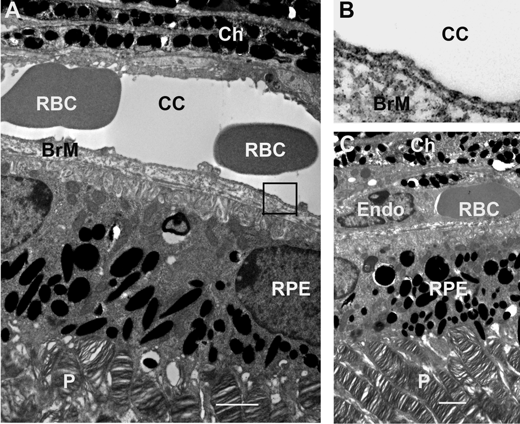
Figure 6.
TEM analysis of the outer retina and choroid from 1-month-old conditional VEGF KO mice with dox induction at E16-18. A, C: Outer retina and choroid. B: Enlarged boxed area in A showing fenestrations. The scale bars in A and C equal to 2 µm. Ch: choroid; RBC: red blood cells; cc: choriocapillaris; BrM: Bruch’s membrane; Endo; endothelial cells; P: photoreceptors. Disruption of RPE-derived VEGF after E15 did not result in detectable ultrastructural changes in choroid, fenestration, RPE, and photoreceptors in mice.

