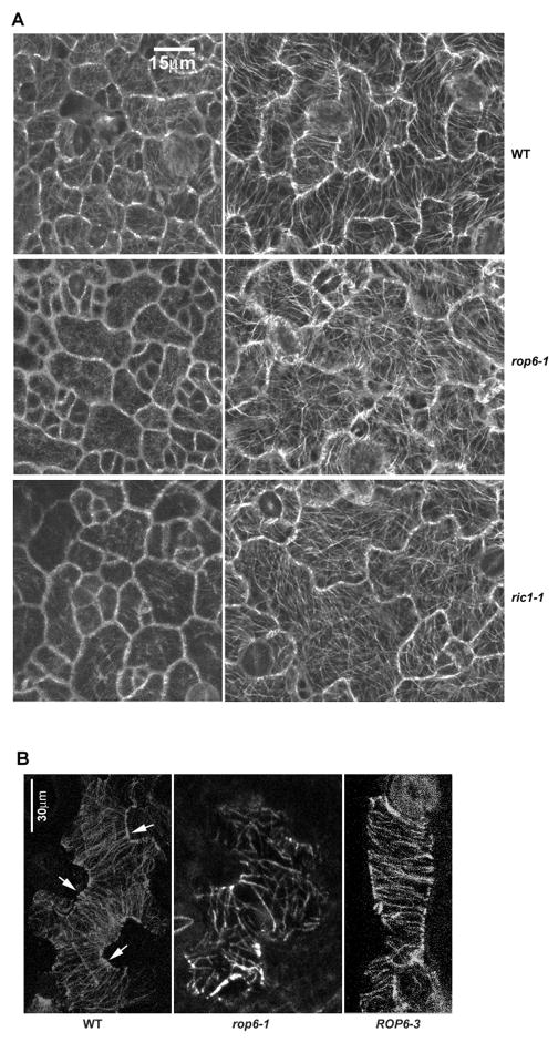
Figure 2. ROP6 promotes the organization of cortical MTs into highly ordered parallel arrays in pavement cells.
(A) Cortical MTs in pavement cells in late stage I (left panel) and late stage II (right panel) from wild type (WT), rop6-1, and ricl-1. MTs were visualized by use of stably expressed tubulin-GFP [17]. MTs in rop6-1 and ric1-1 cells were less ordered and bundled than that in wild-type cells.
(B) Immunolocalization of cortical MTs by use of anti-tubulin antibody confirms rop6-1 cells with more randomly arranged cortical MTs than wild-type cells and that ROP6 overexpression (ROP6-3) promotes the formation of highly ordered transverse MTs aligned perpendicularly to the length of cells. Arrows indicate ordered transverse MTs in the neck regions of WT cells.

