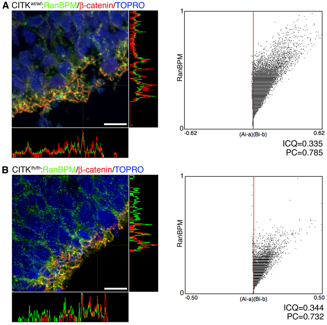
Figure 4. Citron kinase is not necessary for RanBPM expression at the adherens junctions of the VZ surface.
left panels, Endogenous RanBPM co-localizes with β-catenin at the VZ surface of wildtype (A) and CITKfh/fh mutant littermates (B). E17 brain sections from both were immunostained with anti-RanBPM antibody (green) and anti-β-catenin antibody (red). Co-localization of RanBPM and β-catenin is shown at exposed junctional complexes. Orthogonal projections confirming their co-localization show a very similar intensity pattern for the two proteins. Scale bars, 50 µm. right panels, Co-localization analysis with JACoP. Intensity correlation analysis (ICA) was performed for co-localization of RanBPM and β-catenin. A (CITKwt/wt) and B (CITKfh/fh) show intensity correlation plots of the images with the respective plots of the pixel intensities of RanBPM staining (y axis) against their (Ai-a)(Bi-b) values (Ai: individual green (RanBPM) pixel intensity, a: mean of green (RanBPM) pixel intensity, Bi: corresponding red (β-catenin) pixel intensity, and b: mean of red (β-catenin) pixel intensity). Both plots show different intensity co-localization, indicating that absence of CITK does not produce a change in RanBPM localization at the apical VZ junction. ICQ, Intensity correlation quotient value; PC, Pearson’s coefficient.

