
Figure 6.
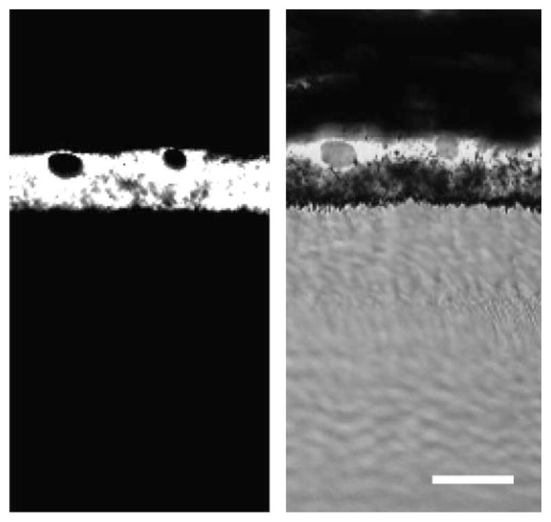
Light microscopic immunohistochemistry of RPE65 antibody in A. ansorgei retina. RPE65 immunoreactivity was intense and specific within the RPE (A) and did not label other regions of the retina (B). Scale bar, 40 μm.
Official websites use .gov
A
.gov website belongs to an official
government organization in the United States.
Secure .gov websites use HTTPS
A lock (
) or https:// means you've safely
connected to the .gov website. Share sensitive
information only on official, secure websites.
Light microscopic immunohistochemistry of RPE65 antibody in A. ansorgei retina. RPE65 immunoreactivity was intense and specific within the RPE (A) and did not label other regions of the retina (B). Scale bar, 40 μm.