
Figure 8.
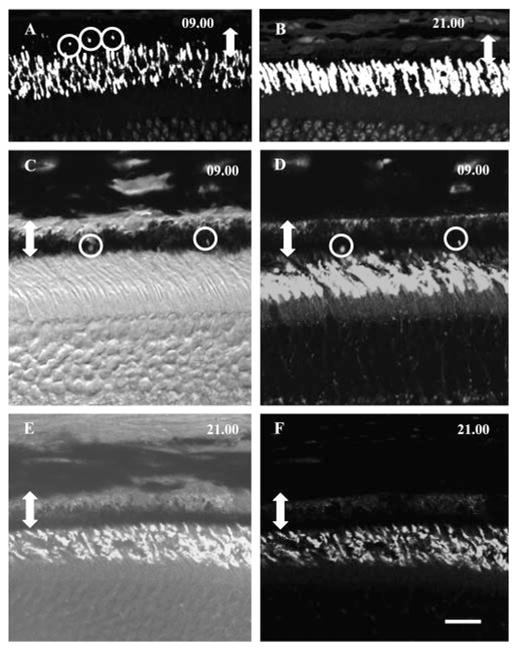
Immunohistochemical detection of rhythmic phagocytosis in rods (A, B) and cones (C–F) of adult A. ansorgei. Sections taken from subjects killed at 0900 (A) and 2100 hours (B) (1 hour after lights on and lights off, respectively) and immunolabeled with rho-4D2 show the presence of numerous immunoreactive inclusions within the RPE (circles) at 0900 but not 2100 hours. Double-headed arrow: the width of the RPE. Sections taken from individuals killed at 0900 (C, D) and 2100 hours (E, F), 1 hour after lights on and lights off, respectively, and immunolabeled with MW-cone opsin showed the presence of immunoreactive inclusions at 0900 but not 2100 hours. (C, E) Merged fluorescence and bright-field images to show the position of immunoreactive phagosomes within the RPE. Double-headed arrow: width of the RPE. Scale bar, 15 μm.