
Fig. 6.
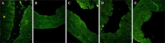
Immunofluorescent stained distal colonic tissues sections in normal mice (a), DSS + PBS treated mice (b) and DSS + rIL-25 treated mice at the dose of 0.2 μg (c), 0.4 μg (d) and 0.8 μg (e), respectively. IL-25 was expressed on the colonic mucosal epithelium surface in normal mice (a) and DSS + 0.4 μg rIL-25 treated mice (d). rIL-25, at indicated doses, or PBS were injected i.p. repeatedly after every 24 h for the duration of 4 days. Mice were killed at day 6