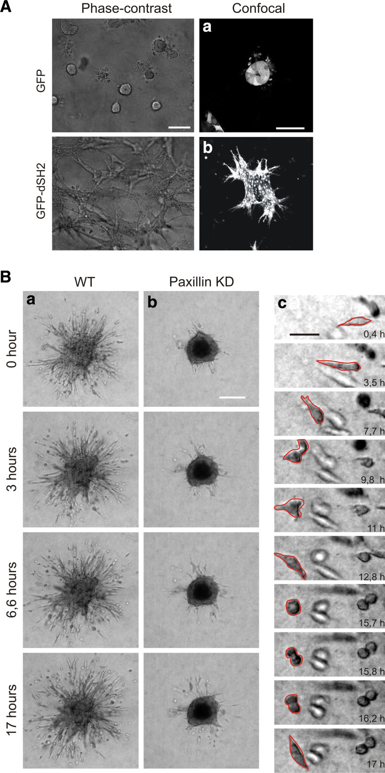
Fig. 5.
Imaging adhesion and cell migration in 3D culture system in vitro. a Phase contrast (scale bar 100 μm) and confocal pictures (scale bar 50 μm) of tubulogenesis assays conducted with LLC-PK1 cells overexpressing either GFP alone (a) or GFP-dSH2 (b) in Matrigel-collagen gels. b Time lapse series (of 17 h) of 4T1 mouse mammary carcinoma cells control (a) and paxillin knockdown (b) invading 3D collagen gels (made with the help of H. Truong). Scale bar 200 μm. c Detailed time lapse serie of one migrating 4T1 control cell. Scale bar 50 μm

