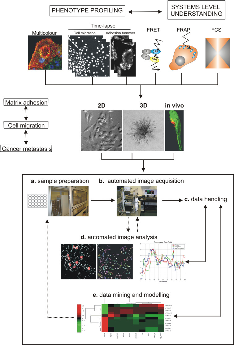
Fig. 7.
Steps for high content systems microscopy approach to understand cancer metastasis. Overview of imaging techniques that allow the phenotypic profiling of proteomics and cellomics (fixed multicolour and time-lapse) and the understanding at systems level (FRET, FRAP and FCS). To enhance our understanding of cancer metastasis, the high-throughput fluorescence microscopy should be applied in 2D, 3D and finally in vivo. a Sample preparation including cell transfection, exposure or immunostaining is nowadays conducted in multi-well dishes using robotics. b Automated image acquisition of fixed or living cells is done using automated microscopes (see Table 1, Available High Content Screening (HCS) instrumentation in [157]). Images can be acquired using different fluorescence microscopy techniques (e.g. fixed multicolour, time-lapse, FRET, FRAP, FCS. c Image data storage requires specialised software and hardware for data handling. d Automated image analysis which needs to be adapted or developed for each assay and is currently a challenge in HTS field. e Another big challenge in the field is the data mining and modelling which requires different disciplines such as statistics and bioinformatics (see Table 2 and available HCS informatics tools in [157]) (adapted from [142])

