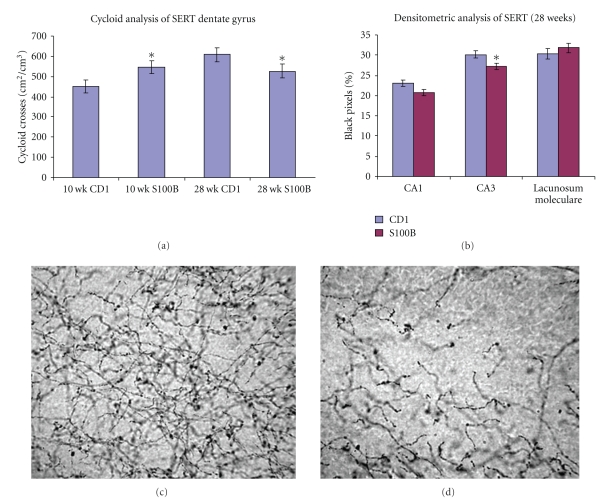
Figure 1.
Serotonergic fiber analysis in the hippocampus of normal and S100B transgenic mice. In (a), a graph of the mean cycloid crosses (cm2/cm3) is shown to indicate the results from the stereological analysis showing a significant increase (P < .05) of SERT fibers in the dentate gyrus of 10-week-old S100B transgenic mice. Alternatively, at 28 weeks, significantly less (P < .05) SERT fibers were observed in the dentate gyrus of S100B transgenic mice. In (b), the results from the densitometric analysis are shown in areas CA1, CA3, and Lacunosum Moleculare of the hippocampus from 28-week-old mice. Note that there is a significant decrease (*P = .027) in the density of serotonergic fibers in the S100B transgenic mice. It is pertinent to note that this densitometric method was verified in the infrapyramidal blade, where the results were consistent with the stereological analysis. There were no significant differences at the 10 week timepoint (data not shown) in CA1, CA3, or Lacunosum Moleculare. In (c) and (d), representative photomicrographs from the infrapyramidal blade of 28-week-old control (c) and S100B transgenic (d) mice. Note the apparent decreased density of serotonergic fibers in the S100B transgenic mice (d).

