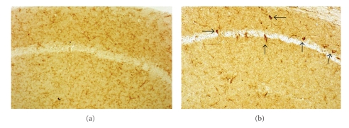
Figure 7.
F4/80-labeling of macrophage/microglia in the CA1 pyramidal cell layer of 1-yr-old CD-1 and S100B transgenic mice. In CD-1 control animals (a), the labeling is found predominantly in resting microglial cells. Alternatively, in the S100B animals (b), several intensely labeled cells (arrows) are observed. These cells could be activated microglia, or another type of immune cell that may have infiltrated the hippocampus, such as macrophages or dendritic cells. 200X.

