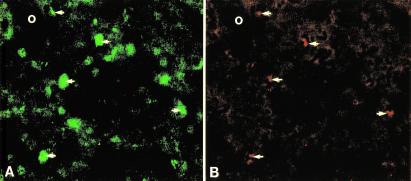
Figure 6.
Double immunofluorescence for CA150 (FITC) (A) and ubiquitin (tetramethylrhodamine B isothiocyanate) (B) immunoreactivities in the same tissue section of caudate nucleus of a patient with grade 3 HD. Circles represent a fiduciary mark. Almost all ubiquitin-positive aggregates (B) (arrows) colocalized with intense staining in puncta in CA150-positive neurons (A) (arrows).

