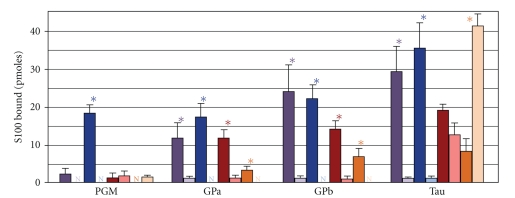
Figure 9.
Interaction of wild-type and mutant S100s with target proteins. Membranes containing 50 pmoles glycogen phosphorylase a (Gpa), glycogen phosphorylase b (Gpb), phosphoglucomutase (PGM), and tau were incubated in 100 nM S100B-488 (blue bars), S100A1-488 (red bars), chimeric S100B-A1-B-488 (purple bars), or S100A1(F88/89A-W90A)-488 (orange bars) in the presence (darker bars) or absence (lighter bars) of Ca2+. The histograms depict that the mean pmoles S100 bound ± the SEM and N's denote no detectable binding. The asterisks denote P ≤ .05, and the N's denote no detectable binding between the ±Ca2+ conditions.

