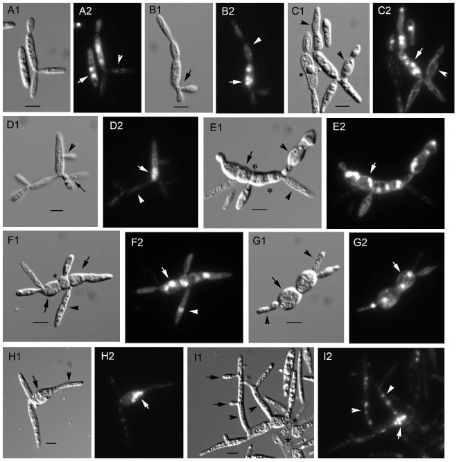
Fig. 3.
Nuclear position and migration in U. maydis lis1-depleted cells. Aliquots of asynchronous exponentially growing cultures of Pcrg1∷lis1 were examined at various times after shift from arabinose to glucose medium. Nuclear position was ascertained by staining with DAPI; cell morphology was analyzed with Nomarski optics. A1–I1. Nomarski view of the DAPI-stained cells in A2–I2, respectively. See text for description of what arrows, arrowheads and asterisks indicate. Bars = 5 μm.

