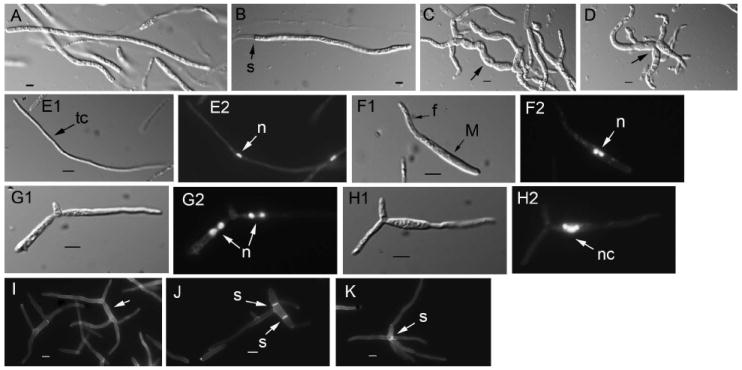
Fig. 8.

Cell morphology of filamentous structures in U. maydis wild type and lis1-depleted cells. A, B. Wild type filaments. Similar structures are formed by Pcrg1∷lis1 strains grown on charcoal arabinose medium. C, D. Filament-like structures formed by lis1-depleted cells. Arrows point to the wider, curved filament-like structures. Samples for the analysis in A–D were from a plate similar to that in Fig. 7. E–K. Strains were grown under nitrogen-limiting conditions. E. Wild type filament consisting of a long tip cell (E1, arrow) with a single nucleus (E2, arrow) (the strain is haploid but able to form filaments). E1. Nomarski view of DAPI-stained cell in E2. F, G and H. Filament-like structures formed by Pcrg1∷lis1 strains. F1. Initiation of filament formation. The mother cell contains two nuclei (F2 arrow). G and H show filament-like structures containing clusters of two nuclei in each appendage (G2 arrows) or multiple nuclei (H2 arrow) in an enlarged cell that gives rise to a filament-like structure devoid of nuclei. E1–H1. Nomarski view of the DAPI-stained cells in E2-H2, respectively. I, J and K. Congo red staining to reveal cell wall and septa. I. Multiple filament-like structures emanating from a central cell (arrow) that lacks septa. J. A mother cell with a filament-like structure and two septa (arrows) at right angles to the long axis of the cell. K. Filament-like structures with a septum (arrow) at their convergence. Bars = 5 μm.
